
Research
Investigating the role of IL34 signaling on mouse brain and behavior.
In my second semester after joining the lab, I became increasingly interested in how neurons and microglia communicate with one another in development. This led me to the colony stimulating factor 1 receptor signaling pathway on microglia. This receptor is expressed on all macrophages in the body, but has a secondary ligand in the brain, that is expressed primarily by neurons (interleukin-34). Existing literature suggested that microglia depend on IL34 signaling in adulthood, but not embryonically, and little was known about when IL34 signaling became relevant in the brain. With my interest in 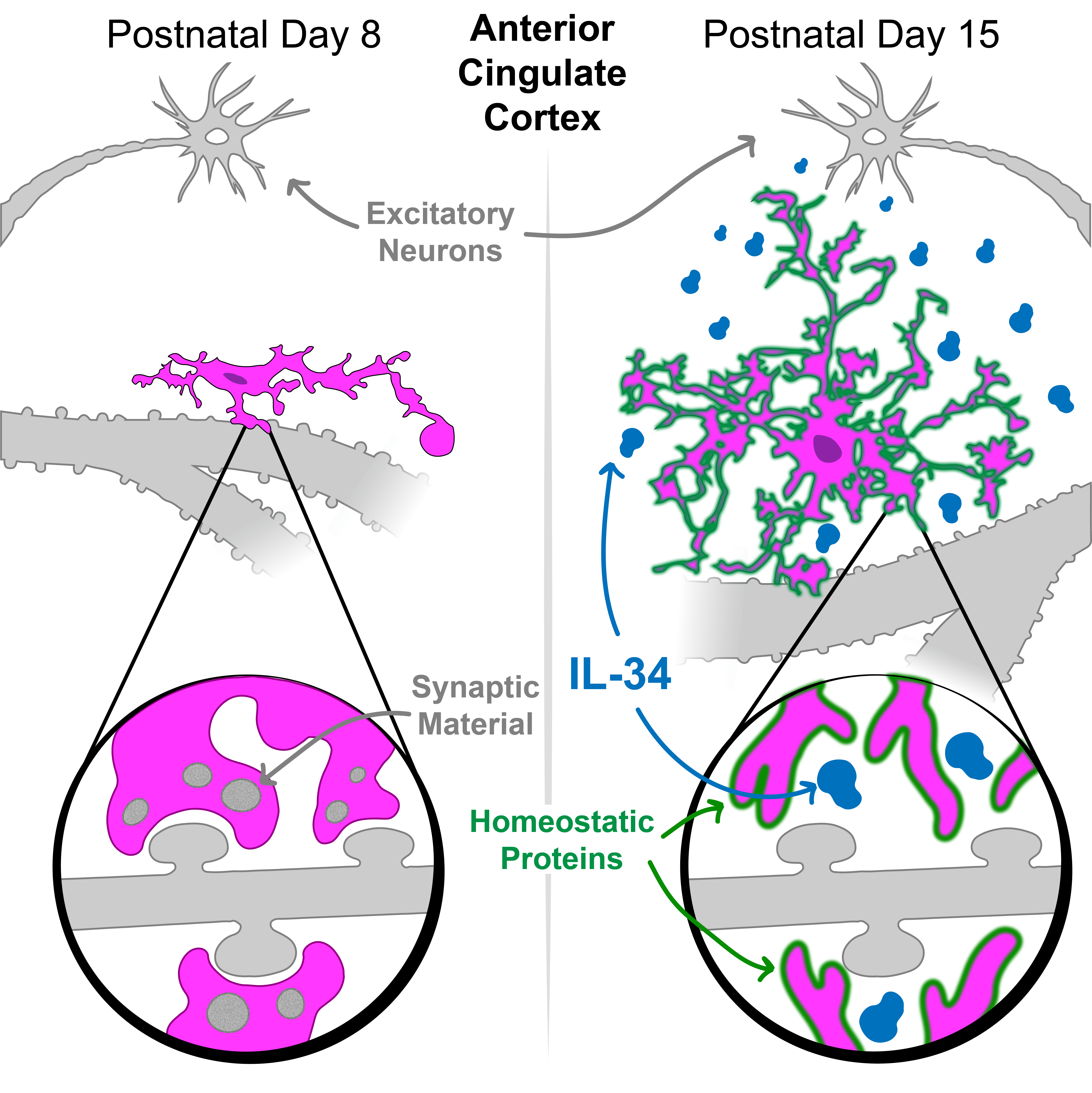 development, I performed a qPCR time course experiment that demonstrated that IL34 expression increases between postnatal day 7 and 15 in mice, a time frame in development when microglia are interacting extensively with neurons across the brain. In collaboration with the lab of Dr. Daniel Saban here at Duke, I obtained mice lacking functional IL34 protein and showed that the microglia in those mice have decreased expression of microglial maturity marker TMEM119, and these mice display deficits in ultrasonic vocalizations. I have presented these findings at both local and national conferences over the past 2 years1,2.
(2025) Excitatory-neuron-derived interleukin-34 supports cortical developmental microglia function. Immunity Paper Link
(2024) Glial Control of Cortical Neuronal Circuit Maturation and Plasticity. Journal of Neuroscience Paper Link
Environmental exposures, development, and microglia
Since joining the Bilbo lab in the fall semester of 2019, I have had the opportunity to collaborate on several projects in the lab investigating the intersection between environmental exposures and development, with a special focus on how microglia translate early-life challenges to later life changes in brain and behavior. The first experiments I was involved with in the lab were with a postdoc (Dr. Alexis Ceasrine) where we found that maternal high-fat diet induces inflammation-dependent, sex-specific behavioral abnormalities in offspring. In males, maternal high fat diet decreased sucrose preference, which was caused by over-engulfment of serotonin neurons by microglia in development and could be rescued with serotonin supplementation. This project has culminated in a paper now available in Nature Metabolism 1. 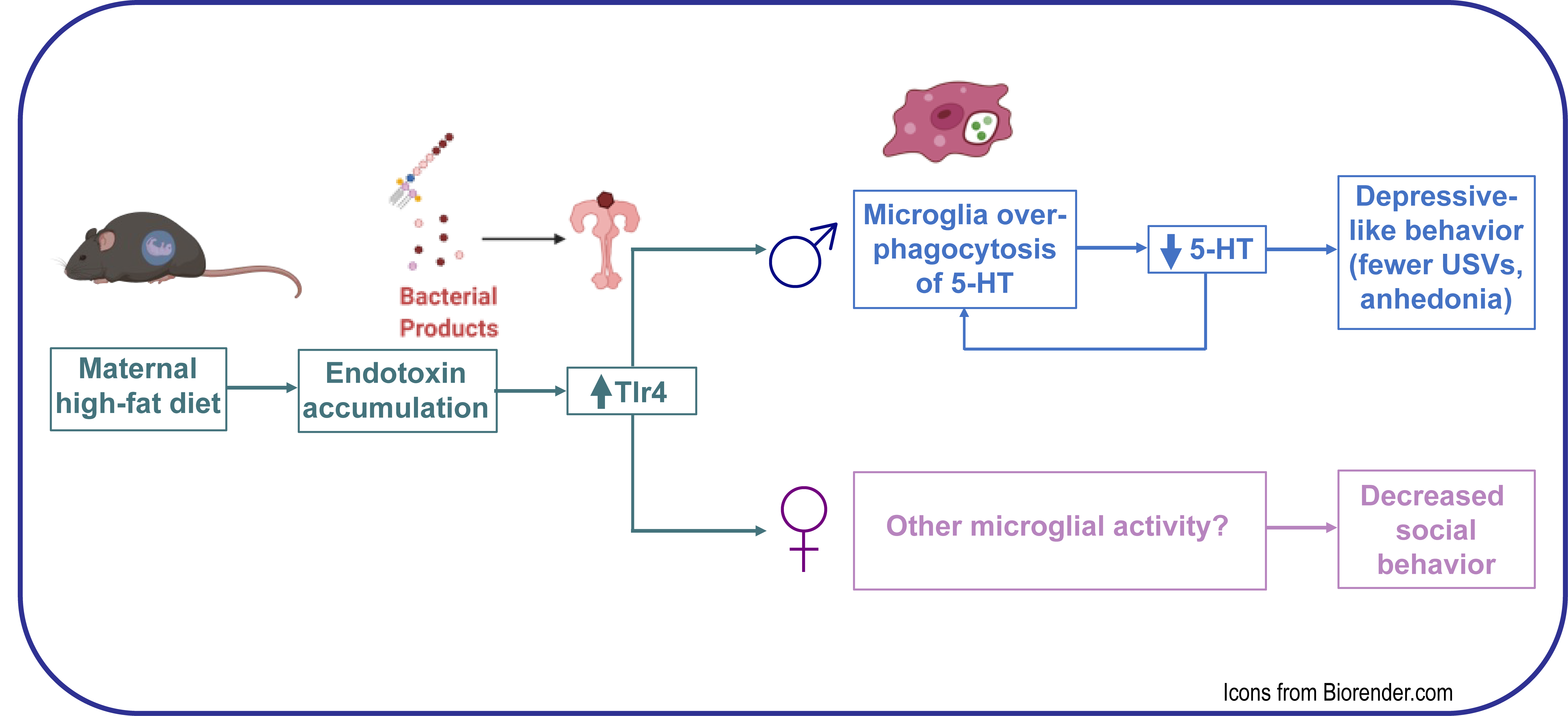 As a follow-up to that project, I also helped with analysis of single cell RNA-sequencing data of isolated microglia and hofbauer cells (placental macrophages) which demonstrated shared origins and transcriptional programs between these two cell types in development2. I have also contributed to a more recent preprint where we found that combined exposure to air pollution and maternal stress in utero causes social deficits in male offspring only. I worked with the postdoc on that project (Dr. Caroline Smith) to analyze bulk RNA-sequencing data from isolated microglia. We found that environmental exposures cause major transcriptional changes in male microglia only, and that microglia following exposure have transcriptional similarities with microglia from germ-free mice. I also helped with DREADD manipulations of dopamine neurons which rescued the social behavior deficits3. While working on these projects I became increasingly interested in microglia and social behavior. Thus, in 2021 I wrote and published a first-author review paper4, where I integrated literature documenting sickness-induced changes in social behaviors across species.
Ceasrine, A. M., Devlin, B. A., Bolton, J. L., Jo, Y. C., Huynh, C., Patrick, B., Washington, K., Joo, F., Campos-Salazar, A. B., Lockshin, E. R., Murphy, S. K., Simmons, L. A., & Bilbo, S. D. (2021). Maternal diet disrupts the placenta-brain axis in a sex-specific manner. Nature Metabolism Paper Link
Ceasrine, A. M., Batorsky, R., Shook, L. L., Kislal, S., Bordt, E. A., Devlin, B. A., Perlis, R. H., Slonim, D. K., Bilbo, S. D., & Edlow, A. G. (2021). Hofbauer cells and fetal brain microglia share transcriptional profiles and responses to maternal diet-induced obesity. Cell Reports Paper Link
Smith, C. J., Rendina, D. N., Kingsbury, M. A., Malacon, K. E., Nguyen, D. M., Tran, J. J., Devlin, B. A., Clark, M. J., Raju, R. M., Burgett, L., Zhang, J. H., Cetinbas, M., Sadreyev, R. I., Chen, K., Iyer, M. S., & Bilbo, S. D. (2022). Microbial modulation via cross-fostering prevents the effects of pervasive environmental stressors on microglia and social behavior, but not the dopamine system. Molecular Psychiatry Paper Link
Devlin, B. A., Smith, C. J., & Bilbo, S. D. (2021). Sickness and the Social Brain: How the Immune System Regulates Behavior across Species. Brain, Behavior and Evolution Paper Link
Investigating the cortical basis of sensory association in autism
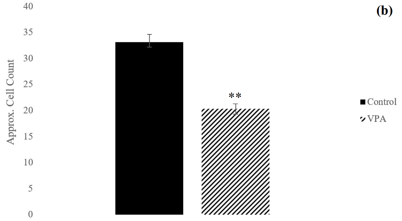 In my undergraduate research experiences, I worked in two labs at the University of Pittsburgh over the summer, and a joint lab at Allegheny College. In the summer after my junior year, I applied for a research fund to facilitate an independent research opportunity in the lab of Dr. Caroline Runyan at Pitt. While there, I worked directly with Dr. Runyan and a new graduate student to train mice on virtual reality behavioral tasks designed to test sensory association (i.e pair a sound with a visual shape). I performed headplate and craniotomy surgeries to image parvalbumin+ interneurons using two-photon calcium imaging while the mice performed the virtual reality behavioral tasks1. I integrated this interest in PV+ interneurons and sensory association with my interest in brain development for my senior thesis research in the lab of Dr. Jeffrey Hollerman. In this project, I found that prenatal exposure to an environmental toxicant (valproic acid) does not influence sensory association ability but decreases the number of PV+ interneurons in adult male rats2. Following this work, I developed and documented a comprehensive protocol for immunostaining, a technique that was new to Dr. Hollerman’s lab.
Devlin, B., Runyan, C. (2018) Summer Research Experience at Pitt: Investigating the Cortical Basis of Sensory Associations. Neuroscience Club Meeting, Meadville, PA. (invited talk)
Devlin, B., Hollerman, J. (2019) Sensory Association and Inhibitory Interneurons in the VPA Rodent Model of